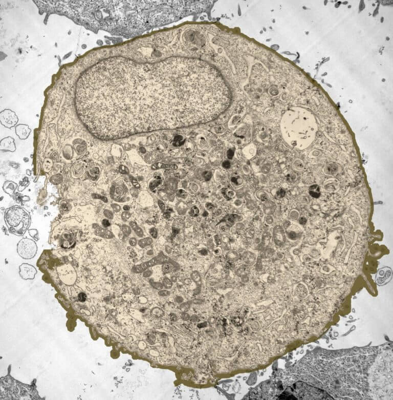
animal cell under electron microscope
4-10 x 10 6 cellsmL WBC. The animal cell is more fluid or elastic or malleable in structure.

Eukaryotic Cells Under The Microscope 2 1 6 Ocr A Level Biology Revision Notes 2017 Save My Exams
A capability for scanning electron microscopy of wet biological specimens is presented.

. Below the basic structure is shown in the same animal cell on the left viewed with the light microscope and on the right with the transmission electron. Hemocytometer protocol filling sheet. 4-6 x 10 9 cellsmL RBC.
Animal Cell Under An Electron Microscope. Animal cells have a basic structure. Both plant and animal cells comprise membrane-bound organelles such.
Light and electron microscopes allow us to see inside cells. May 4 2017 - cells under electron microscope - Google Search. Cells range in size.
Once the mouse is placed under the microscope scan the tissue using the eyepiece for the region of interest. Plant animal and bacterial cells have smaller components each with a specific function. The electron microscope Electron microscopes use a beam of electrons instead of beams or rays of light.
Animal cell under electron microscope. Use the endogenous fluorescence of transgenic immune cells or tumor cells. Up to 20 cash back Stock photography Electron microscope images of animal cells with nucleus and organelles 353379966 Download pictures from the photo stock library.
The present study was undertaken to show the applicability to biological studies of a scanning electron microscope SEM equipped with a high-sensitive cathodoluminescence detection. The animal cell is more fluid or elastic or malleable in structure. How would you identify if a cell was a plant or animal cell when looking at it under a microscope.
Observing a wide range of biological processes and animal cell under light microscope is easier due to advances in microscopic techniques. Beneath a plant cells cell wall is a cell membrane. May 4 2017 - cells under electron microscope - Google Search.
A membrane that is transparent to electrons protects the fully hydrated sample. Electron Microscopic Study Of Cell And Organelles Important The largest known animal cell is the ostrich egg which can stretch. When autocomplete results are.
Animal cell under electron microscope. Most animal cells are between 001 mm 005 mm. An animal cell also contains a cell membrane to keep all the organelles.
However their presence was inferred long before their observation due to plasmolysis bursting of animal cells placed. Under a microscope plant cells from the same source will have a uniform size. Most cells both animal and plant range in size between 1 and 100.
Because of this dimension they are not visible under optical microscope. An animal cell also contains a cell membrane to keep all the organelles and cytoplasm contained but it lacks a cell wall. If you overdilute you will count less cells so your counts might be more.
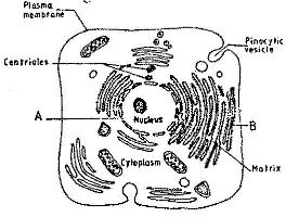
A typical animal cell as seen in an electron microscope Medical Images For PowerPoint 1. Greg Foot explains the main differences between light and electron microscopes. Up to 24 cash back Structure of plant and animal cells under an electron microscope Advanced Higher Biology Cell and molecular Biology The Electron Microscope Two main.
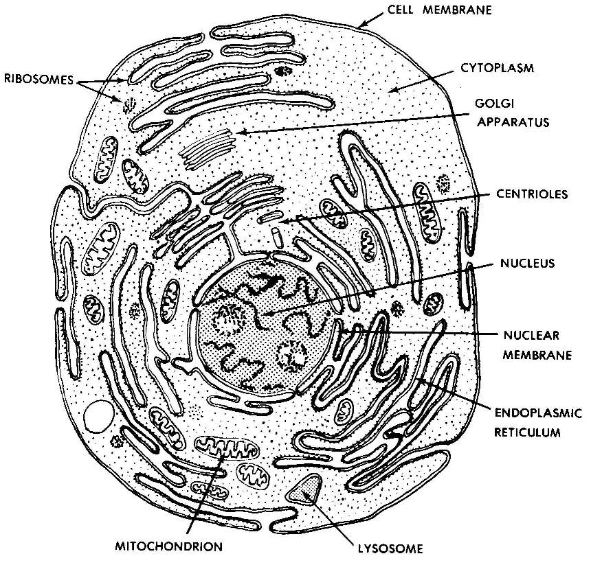
Typical Animal Cell Pinocytotic vesicle Lysosome Golgi vesicles Golgi vesicles.

Electron Microscope Radioautographs Of Profiles Of Liver Cells From Download Scientific Diagram
The Cell Kcse Biology Notes Objectives Syllabus Questions Answers And Others Atika School

Gce Cie Biology Animal And Plant Cell Structures And
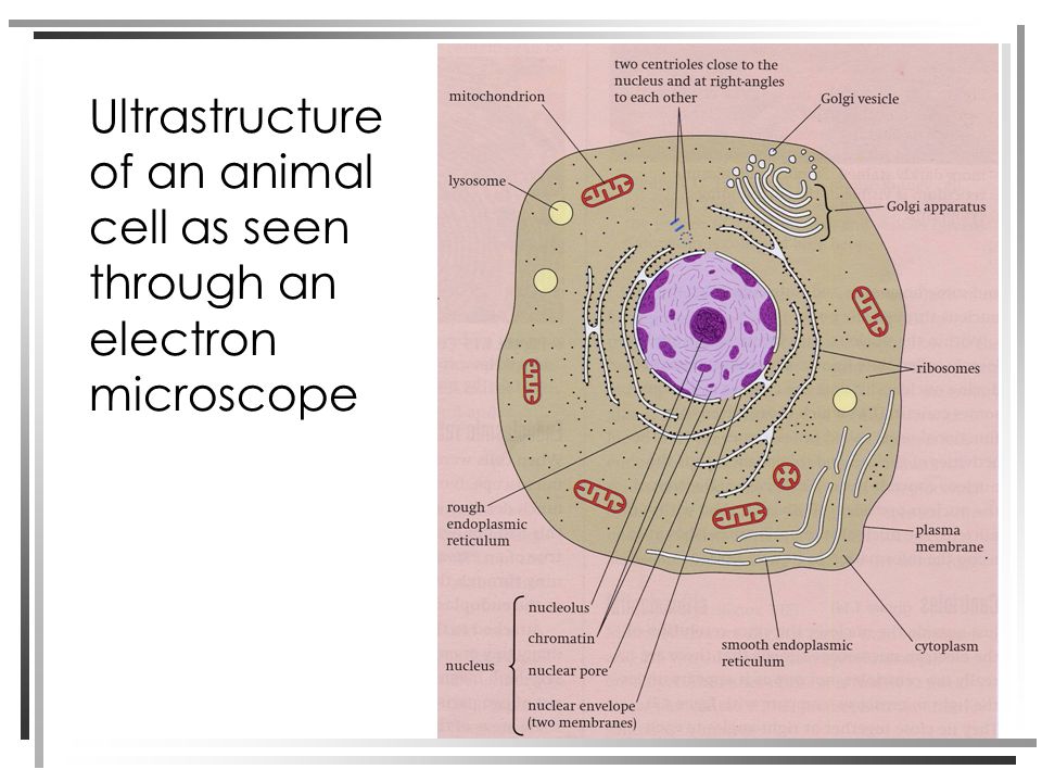
Structure Of Plant And Animal Cells Under An Electron Microscope Ppt Video Online Download

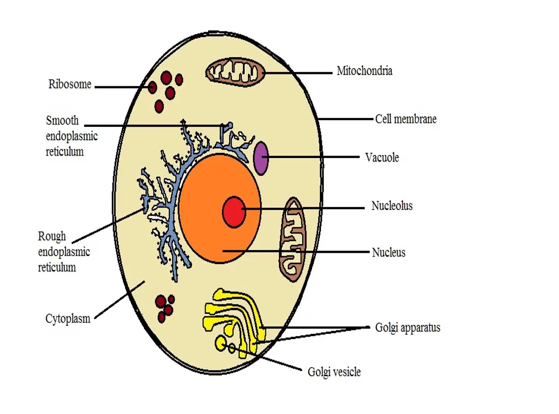
Animal Cell Plant Cell Diagram Cell Diagram Animal Cell
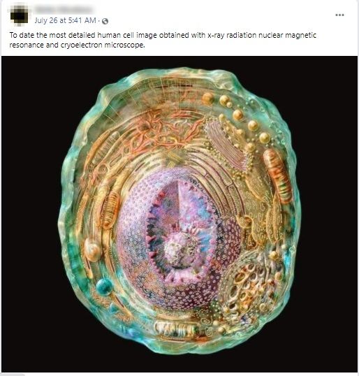
Illustrated Animal Cell Picture Falsely Shared As The Detailed Image Of A Human Cell Factly
Images 01 Introduction And Terminology Basic Human Anatomy

Rana Ray Diagram Of Animal Cell Seen Through Electron Microscope Brainly In
Electron Micrographs

Magnification Questions Doc Cell Magnification Fig 1 2 1 Below Shows An Animal Cell 5m Fig 1 2 1 Diagram Showing The General Structure Of An Course Hero
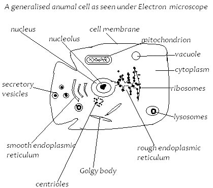
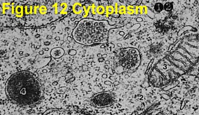
The Figure Below Is A Fine Structure Of A Generalized Animal Cell As Seen Under An Electron Microscope
What Are The Differences Between A Plant Cell And An Animal Cell

Images 01 Introduction And Terminology Basic Human Anatomy

A Typical Animal Cell As Seen In An Electron Microscope Medical Ima
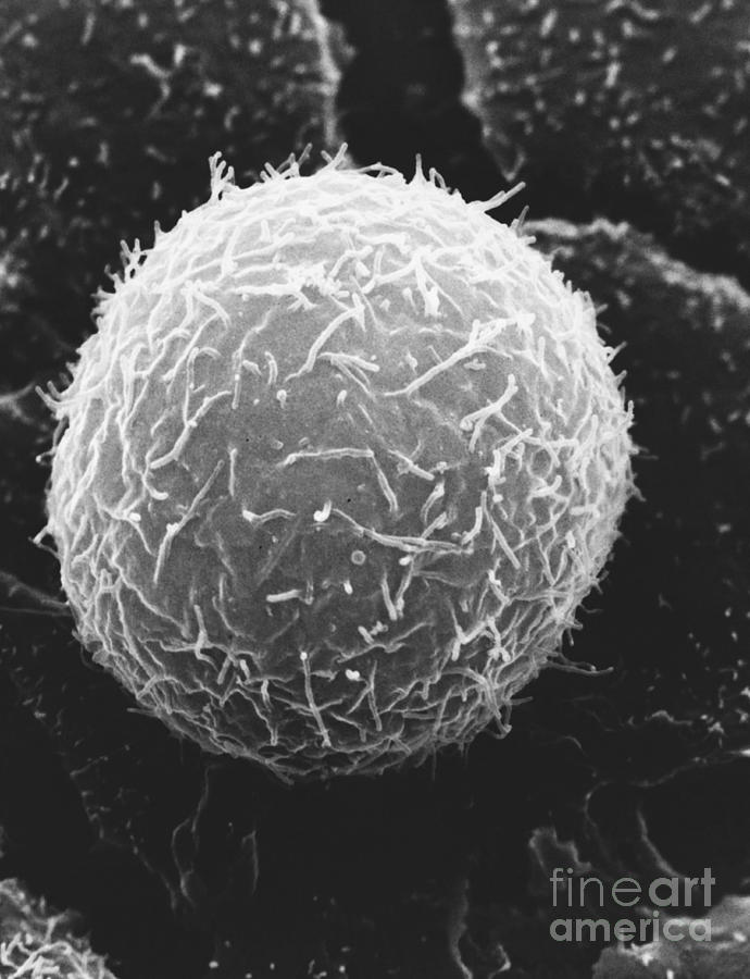
Typical Animal Cell Sem Photograph By David M Phillips Fine Art America

Animal Cell Definition Structure Parts Functions Labeled Diagram
Amazing 27 Things Under The Microscope With Diagrams
![]()
Electron Micrograph Animal Cell Hi Res Stock Photography And Images Alamy
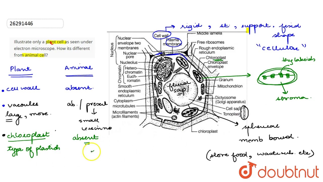
Illustrate Only A Plent Cell As Seen Under Electron Microscope How Its Different From Animal Cell Youtube